Examqo › Examqo library › Speech-language pathology and audiology › Hearing science
Cochlear physiology: travelling wave, hair cells, potentials and the cochlear amplifier
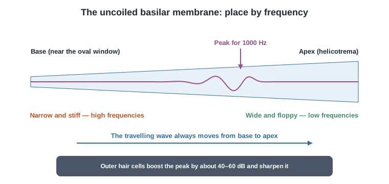
The basilar membrane sorts sound by frequency along the cochlea; the inner hair cells are the sensors that send information to the nerve, and the outer hair cells are an active amplifier that adds sensitivity and sharp tuning. Most cochlear losses start with the loss of this amplifier.
Why it matters in the exam
The bank's anatomy questions are simple but exact: the upper chamber is the scala vestibuli (perilymph, starting at the oval window), the middle is the scala media (potassium-rich endolymph, a potential of about +80 mV generated by the stria vascularis on the outer wall of the duct), and the lower is the scala tympani (perilymph, ending at the round window); Reissner's membrane separates the upper from the middle. A single row holds about 3500 inner hair cells, while about 12,000 outer hair cells form three to four rows; the tectorial membrane grips only the longest outer-cell stereocilia.
The hard questions are about the process. Bending the stereocilia toward the tallest one opens channels, potassium flows in from the endolymph and depolarises the cell — a driving force of about 150 mV (80 minus minus 70). In the inner cell this releases glutamate onto type I nerve fibres; in the outer cell the protein prestin shortens the cell on depolarisation and lengthens it on hyperpolarisation — this electromotility boosts soft sounds by about 40–60 dB and sharpens the tip of the tuning curve. In a dead (cadaver) cochlea a travelling wave appears only at very high levels and is broad. The cochlear microphonic (CM) comes mainly from outer cells, follows the waveform and flips with polarity; the summating potential (SP) is a DC shift to which inner cells contribute most.
Key points
- Base — highs, narrow and stiff; apex — lows, wide and floppy.
- About 3500 inner cells that send the message; about 12,000 outer cells that amplify.
- Endolymph: high potassium, about +80 mV, from the stria vascularis.
- Prestin: depolarisation shortens; the amplifier adds about 40–60 dB and sharp tuning.
- The CM comes from outer cells and flips with polarity; the SP is a DC shift.
Summary table
| Topic | Core fact | Clinical link |
|---|---|---|
| Travelling wave | Moves from base to apex; high frequencies at the base, low at the apex | Tonotopy begins on the basilar membrane |
| Inner hair cells | About 3500, one row; the sensors; about 95% of afferent fibres | Damage — deeper cochlear loss and loss of information |
| Outer hair cells | About 12,000; prestin; cochlear amplifier | Damage — threshold rises up to about 40–50 dB, broad tuning, recruitment, no OAE |
| Endocochlear potential | About +80 mV in the endolymph, from the stria vascularis | A driving force of about 150 mV for the potassium current |
| CM and SP | CM — AC mainly from outer cells; SP — DC shift mainly from inner cells | The CM flips with stimulus polarity; useful in diagnosing ANSD |
| Tuning curve | Sharp tip at the characteristic frequency and a broad tail | Outer-cell damage raises and broadens the tip; the tail stays |
| Ototoxic drugs and noise | Hit the outer cells of the basal turn first | Loss starts in the high and extended high frequencies |
A free exam question
What happens in the electromechanical transduction stage in the cochlea?
- A. An inner hair cell elongates
- B. An outer hair cell contracts
- C. An outer hair cell releases glutamate
- D. An inner hair cell releases acetylcholine
In the full version for subscribers
- A simple algorithm
- Memory aid
- Exam traps
- 5 review cards
- What matters for the exam
The full version is open to subscribers of: Speech and hearing (communication disorders).
Subscriber? Read the full topic Plans and prices 25 free questions
Sources
- Pickles, An Introduction to the Physiology of Hearing, 4th edition (2012) — The cochlea; mechanics and transduction
- Robles & Ruggero, Physiological Reviews, 2001 — Mechanics of the mammalian cochlea
- Dallos, Journal of Neuroscience, 2008 — The active cochlea (prestin and electromotility)
Sources are given for further reading; the text is written afresh and not copied from them.
Reviewed by the Examqo AI Professor · 2026-11-02. The text may contain errors; when in doubt, check the source.