Examqo › Examqo library › Speech-language pathology and audiology › Clinical audiology
Reading the audiogram: types of hearing loss, grades, shapes and tuning fork tests
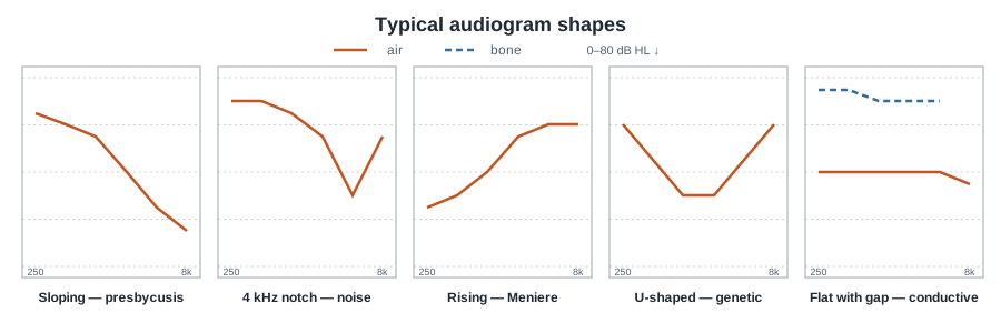
The type of loss is set by the relation between air and bone conduction — an air-bone gap shows a conductive component, equal and impaired thresholds show a sensorineural loss — and the shape of the curve points to the cause: sloping in presbycusis, a 4 kHz notch with noise, rising in Ménière's disease, and a Carhart notch in bone conduction in otosclerosis.
Why it matters in the exam
Questions give an audiogram or a description of one and ask for the type and cause: an air-bone gap shows a conductive component; a sensorineural loss comes from damage to the hair cells or the nerve; a rising audiogram means a greater loss in the low frequencies; a U-shaped audiogram means the low and high frequencies are better than the middle; ototoxic drugs hit the high frequencies first; and presbycusis leads the causes of adult sensorineural loss, with a mild to moderate sloping curve.
Special cases are asked too: a Carhart notch — a bone-conduction dip that is largest at 2,000 Hz — is typical of otosclerosis and is not true sensory damage; an air-bone gap can also arise in the inner ear (a "third window"), for example with superior semicircular canal dehiscence or an enlarged vestibular aqueduct. In tuning fork tests: the Weber is heard in the ear with the conductive loss or away from the ear with the sensorineural loss, and a positive Rinne (air better than bone) shows there is no significant conductive loss.
Key points
- Air-bone gap = conductive component; equal, impaired thresholds = sensorineural.
- Presbycusis — the common cause, sloping.
- Noise notch around 4 kHz; ototoxicity in the high frequencies.
- Carhart notch at 2,000 Hz — otosclerosis.
- Weber to the conductive ear; positive Rinne — no conductive loss.
Summary table
| Type or shape | What it looks like | Typical cause |
|---|---|---|
| Conductive loss | Normal bone thresholds, impaired air thresholds, an air-bone gap | Middle ear fluid, perforation, otosclerosis, ossicular discontinuity |
| Sensorineural loss | Air and bone thresholds equally impaired | Hair cells or nerve: presbycusis, noise, ototoxicity |
| Mixed loss | Bone thresholds impaired and an air-bone gap too | A combination of both mechanisms |
| Sloping / high frequencies | A loss that grows towards the high frequencies | Presbycusis; ototoxicity starts in the high frequencies |
| Noise notch | A dip around 3–6 kHz, mainly 4, with recovery at 8 | Noise exposure |
| Rising, U-shaped and peaked | Rising — worse in the lows; U — the middle is worse; peaked — in the fluctuating stage | Ménière's disease (rising, peaked); genetic (U) |
| Carhart notch and third window | Maximal bone dip at 2,000 Hz; or a gap that arises in the inner ear | Otosclerosis; superior canal dehiscence, enlarged vestibular aqueduct |
| WHO 2021 grades (better-ear average of 0.5–4 kHz) | Mild 20–34, moderate 35–49, moderately severe 50–64, severe 65–79, profound 80–94, complete 95 and above | Normal hearing: below 20 dB |
In the full version for subscribers
- A simple algorithm
- Memory aid
- Exam traps
- 5 review cards
- What matters for the exam
The full version is open to subscribers of: Speech and hearing (communication disorders).
Subscriber? Read the full topic Plans and prices 25 free questions
Sources
- World Health Organization, World Report on Hearing, 2021 — Grades of hearing loss
- Katz (ed.), Handbook of Clinical Audiology, 7th edition (2015) — Interpreting the audiogram; tuning fork tests
- Merchant & Rosowski, Otology & Neurotology, 2008 — Conductive hearing loss caused by third-window lesions of the inner ear
Sources are given for further reading; the text is written afresh and not copied from them.
Reviewed by the Examqo AI Professor · 2026-11-02. The text may contain errors; when in doubt, check the source.